
The Human BioMolecular Atlas Program (HuBMAP)
The Human BioMolecular Atlas Program (HuBMAP)
Image of the Week
Some of the most amazing things to come out of the HuBMAP Consortium are the images of healthy human tissues generated by our researchers.
Here, we collected them in one place to celebrate the work of these talented individuals.
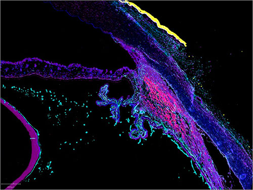
CODEX image of the anterior eye, courtesy of Drs. Angela Kruse and Thai Pham at Vanderbilt University
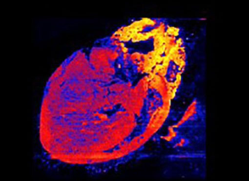
DESI image of the concentration of oleic acid across mouse heart, courtesy of Drs. Liming Pei (CHOP) and Brent Stockwell (Columbia)
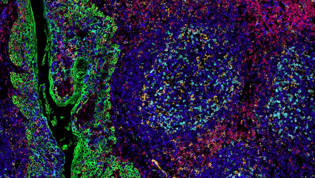
CODEX image of skin, by Dr. Athanasios Ploumakis of the Vlachos lab at Beth Israel Deaconess Medical Center
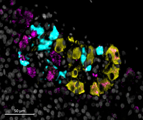
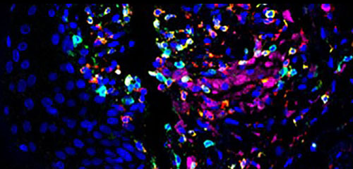
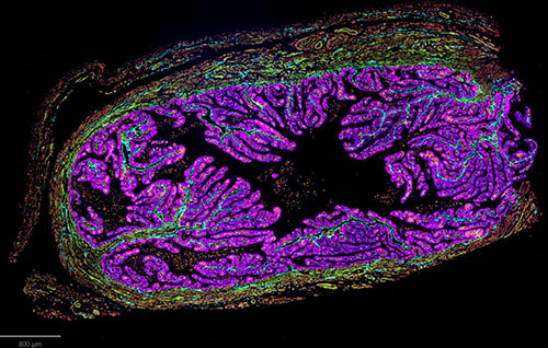
CycIF image of ganglia within the pancreas, courtesy of Sam Ewing at the University of Florida
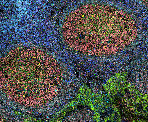
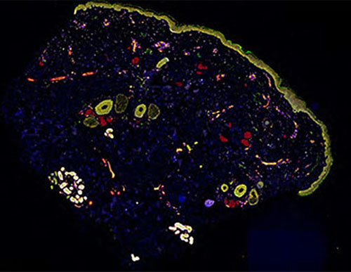
MACSima image of tonsils, courtesy of Drs. Werner Muller and Andreas Bosio at Miltenyi Biotec
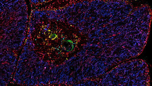
IBEX image of thymus, courtesy of Dr. Andrea Radtke, in the Germain lab at NIAID
Cell DIVE image of skin, courtesy of Liz McDonough of GE Research
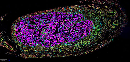
CODEX image of the ampulla from Dr. Kate O'Neill of UPENN
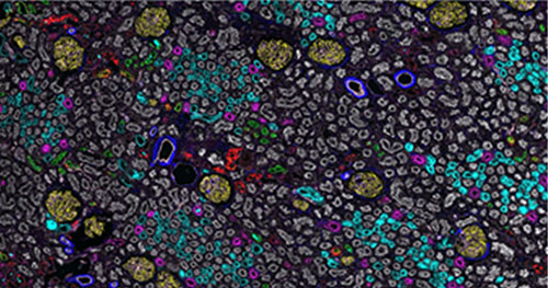
CODEX image of kidney, courtesy of Dr. Tarek Ashkar of Indiana University
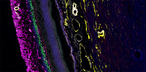
CODEX image of retina, courtesy of Dr. Angela Kruse at Vanderbilt University
IBEX image of a tonsil, courtesy of Drs. Kartika Padhan and Andrea Radtke in the Germain lab at NIAID
CODEX image of the infundibulum, courtesy of Dr. Kate O'Neill from UPENN
