
The Human BioMolecular Atlas Program (HuBMAP)
The Human BioMolecular Atlas Program (HuBMAP)
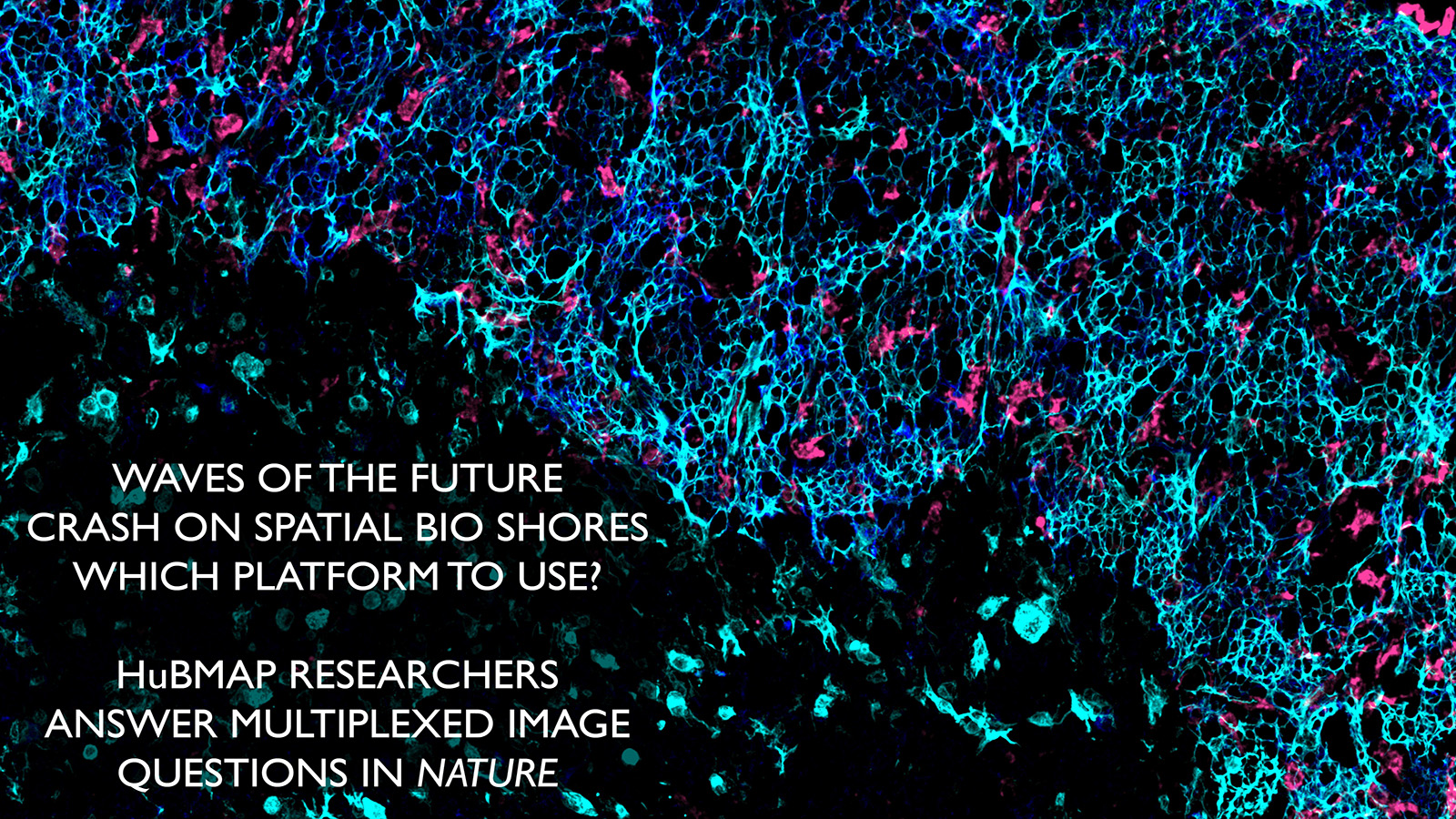
Image of the Week
Some of the most amazing things to come out of the HuBMAP Consortium are the images of healthy human tissues generated by our researchers.
Here, we collected them in one place to celebrate the work of these talented individuals.
CellDIVE of skin courtesy of Drs. Fiona Ginty, Soumya Ghose, and Liz McDonough from GE Research. Special effects by Yingnan Ju at Indiana University
CODEX of intestine from Dr. John Hickey at Stanford
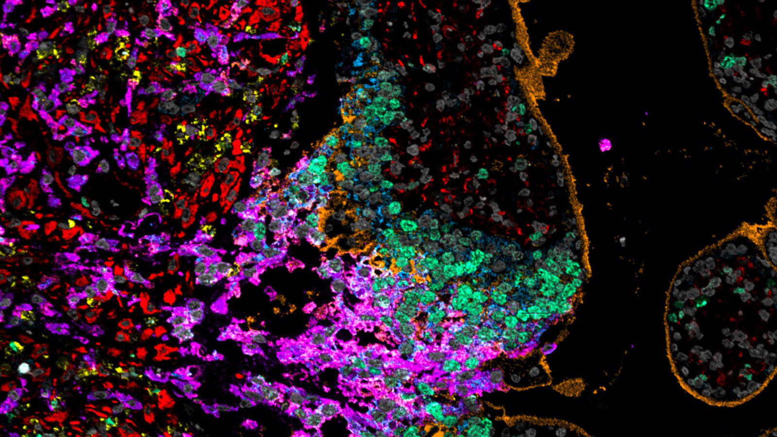
CODEX image of heart ventricles, courtesy of Dr. Kai Tan at CHOP
IBEX image of human thymus, courtesy of Andrea Radtke of the Germain lab at NIAID
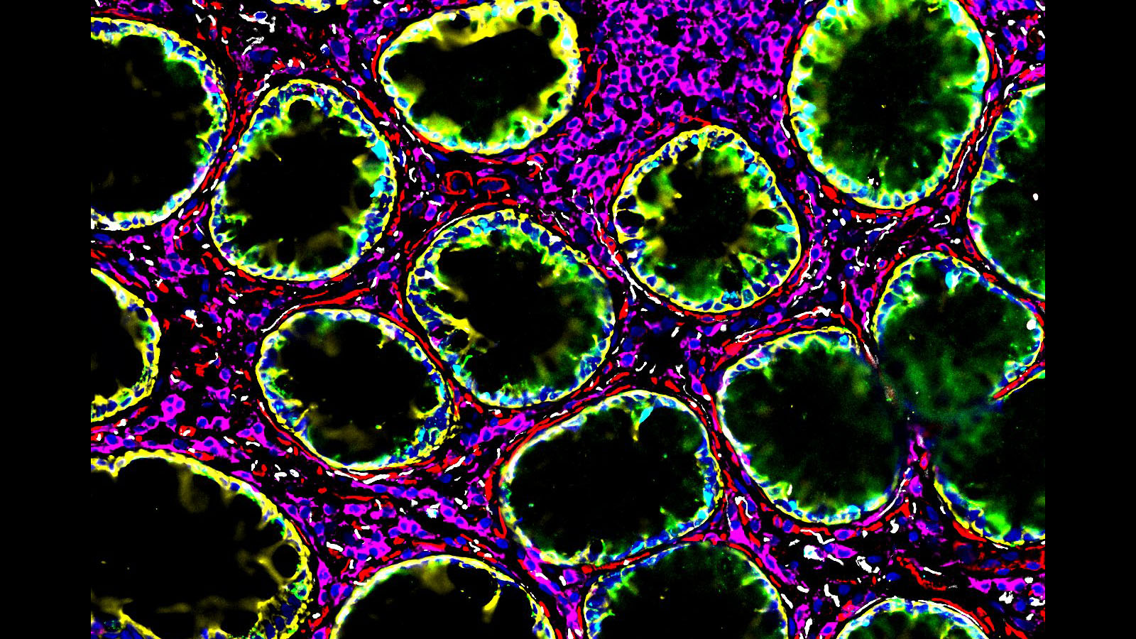
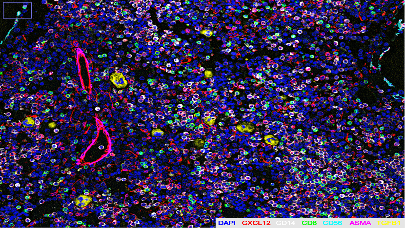
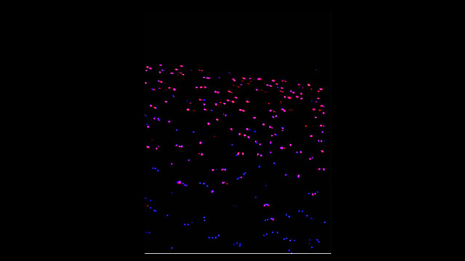
Imaging mass cytometry image of the placenta, courtesy of Santhosh Sivajothi of the Robson lab at JAX
CODEX of bone marrow from sternum, courtesy of Dr. Kai Tan at Children's Hospital of Philadelphia
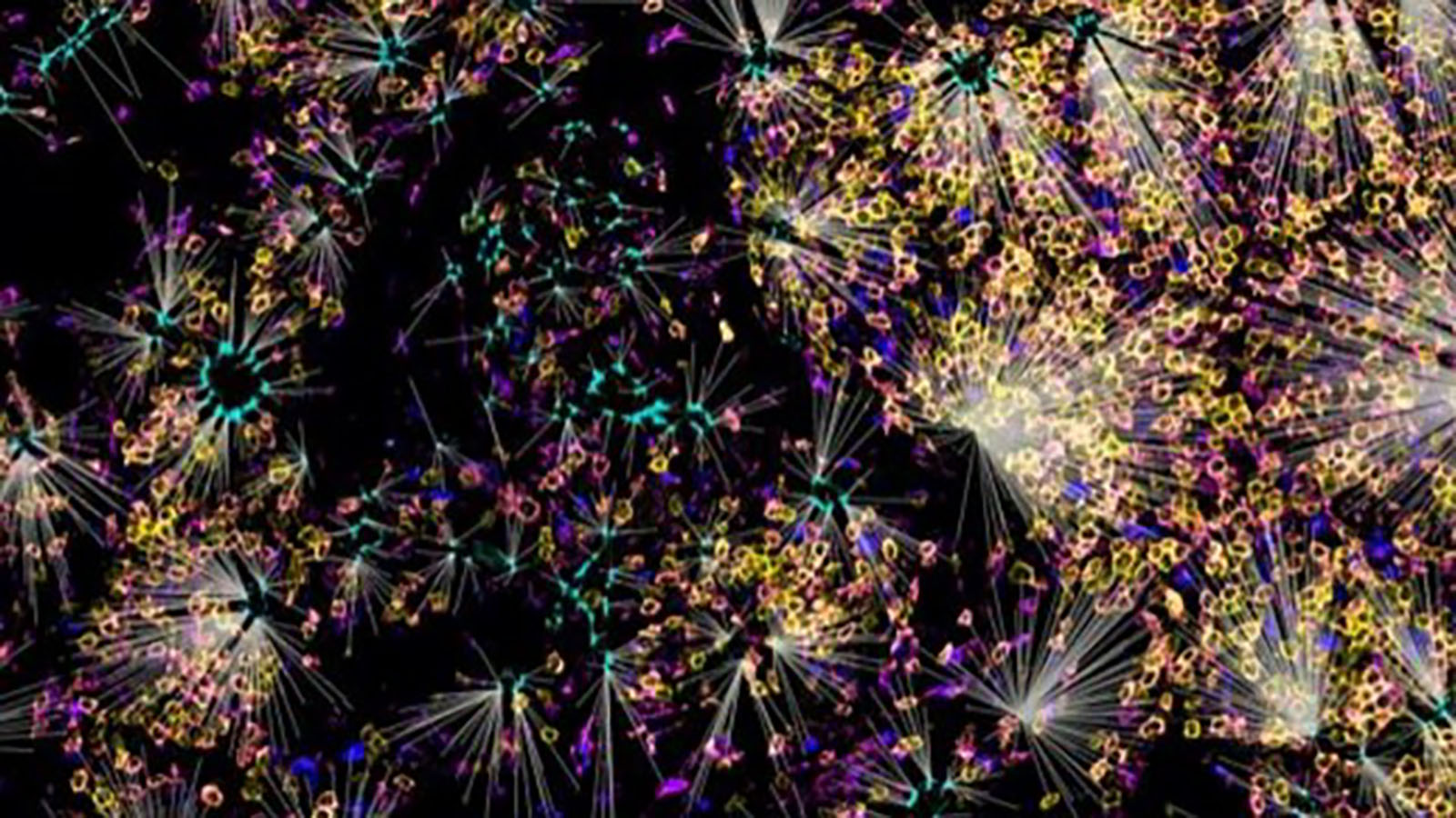
Confocal microscopy image of the human lens courtesy of Angela Kruse
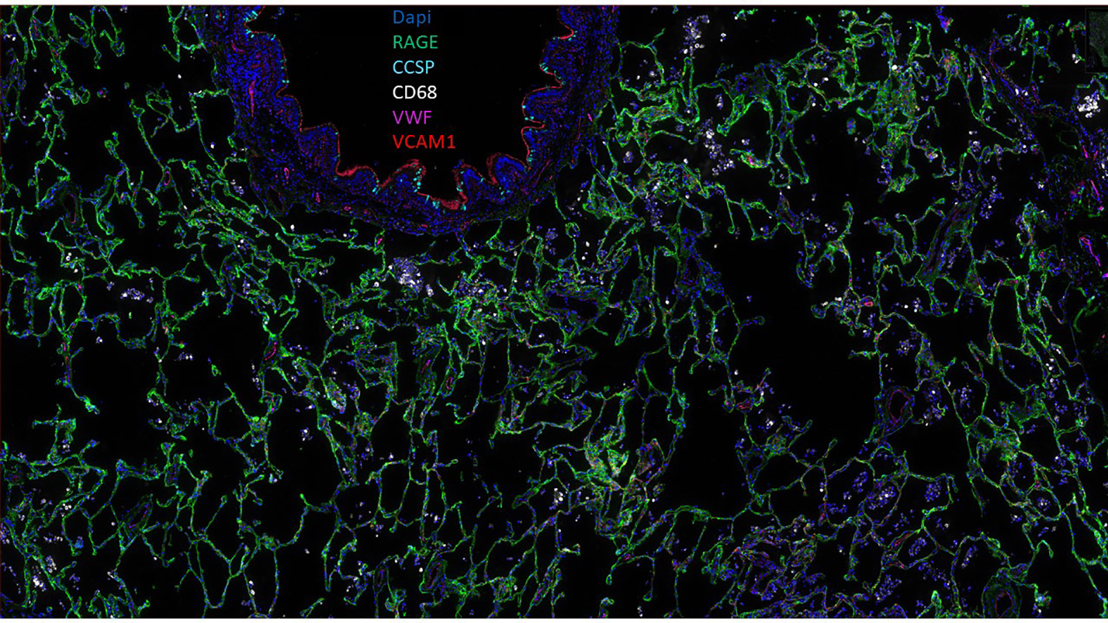
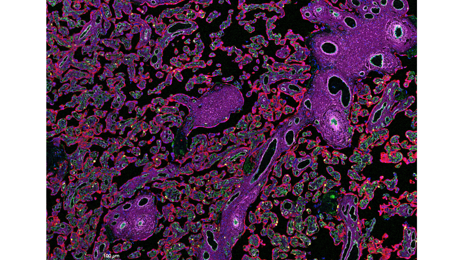
CellDIVE image of healthy human lung courtesy of Gloria Pryhuber, Fiona Ginty, Lisa Lowery, and Christine Surrette
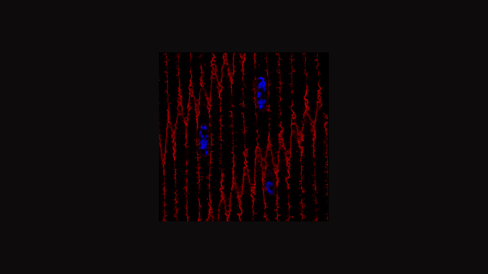
MERFISH image of articular cartilage courtesy of David Rowe's lab at UConn
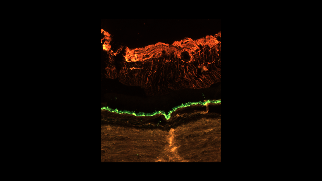
Immunofluoscence microscopy image of a human retina courtesy of Dr. Angela Kruse of Vanderbilt University
MIBITOF image of decidua, courtesy of Mike Angelo lab at Stanford University
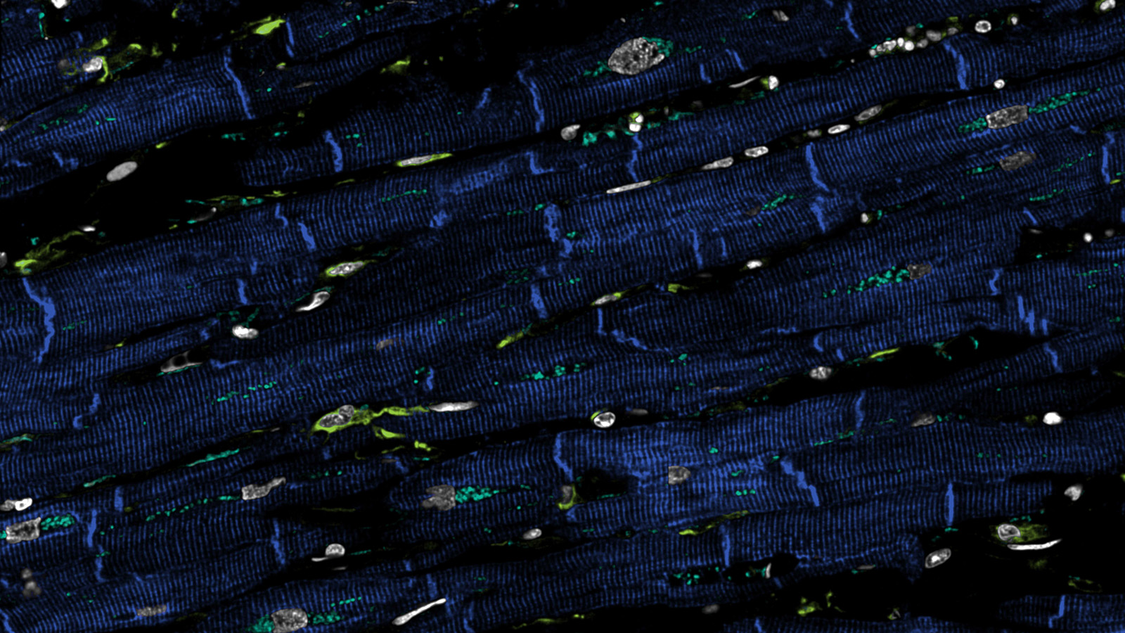
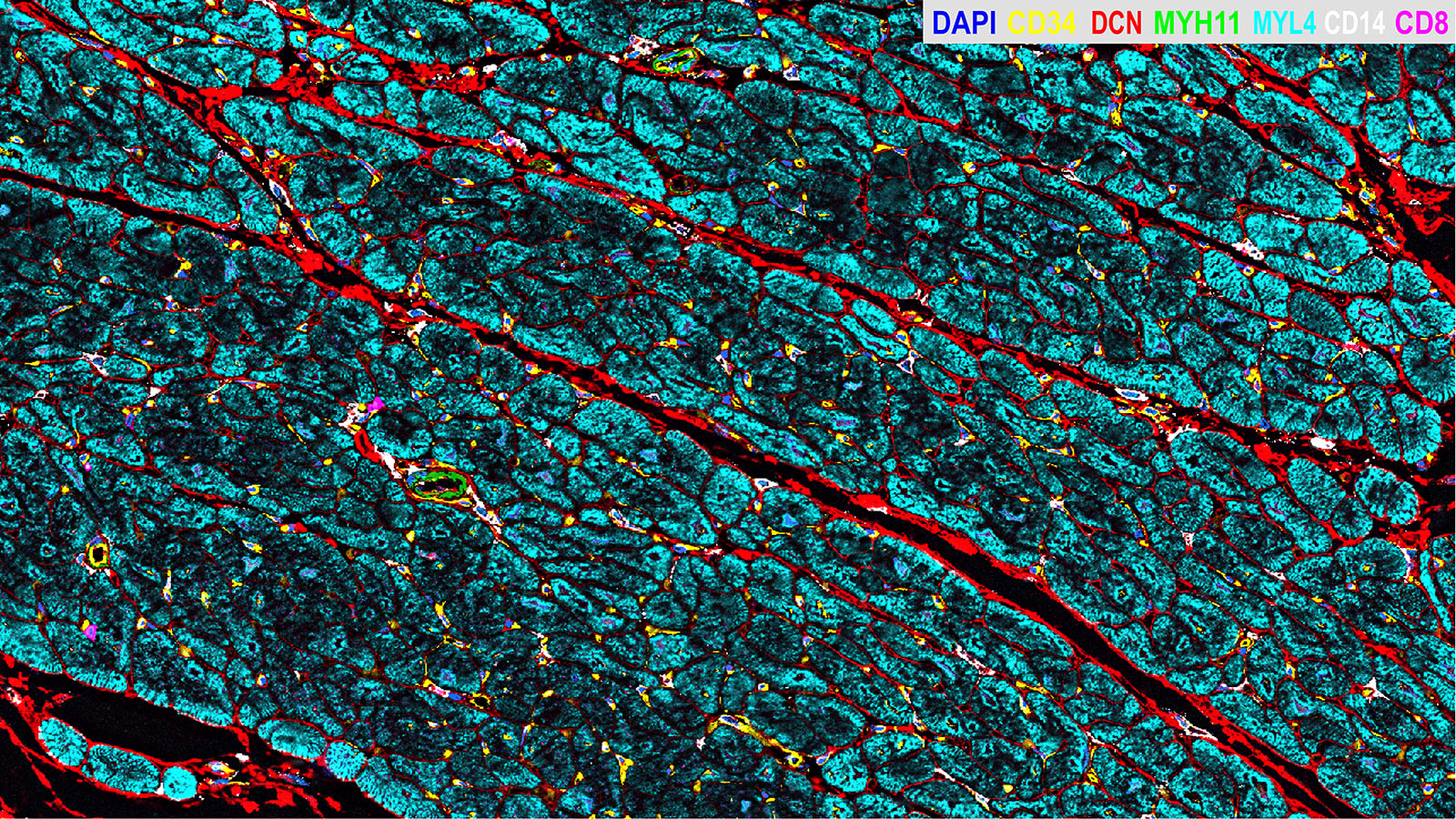
Confocal microscopy image of human heart, courtesy of Dr. Andrea Radtke of the Germain Lab at NIAID
