
The Human BioMolecular Atlas Program (HuBMAP)
The Human BioMolecular Atlas Program (HuBMAP)
Image of the Week
Some of the most amazing things to come out of the HuBMAP Consortium are the images of healthy human tissues generated by our researchers.
Here, we collected them in one place to celebrate the work of these talented individuals.
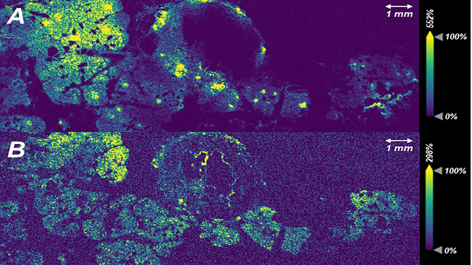
MALDI-IMS image of pancreas courtesy of Kevin Zemaitis of PNNL
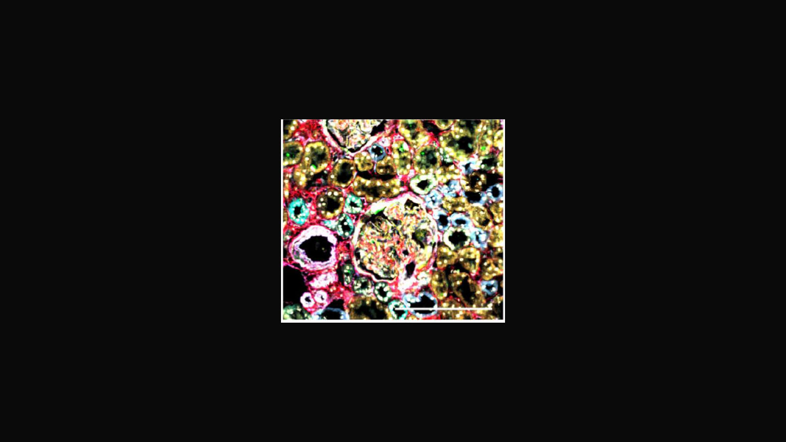
Immuno-SABER image of kidney courtesy of Matt Serrata in Peng Yin's lab at Harvard
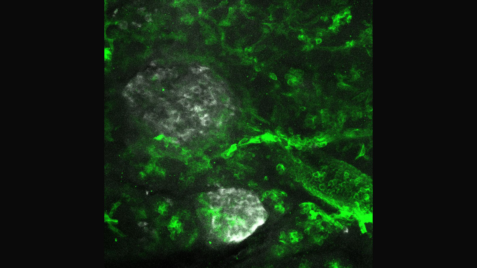
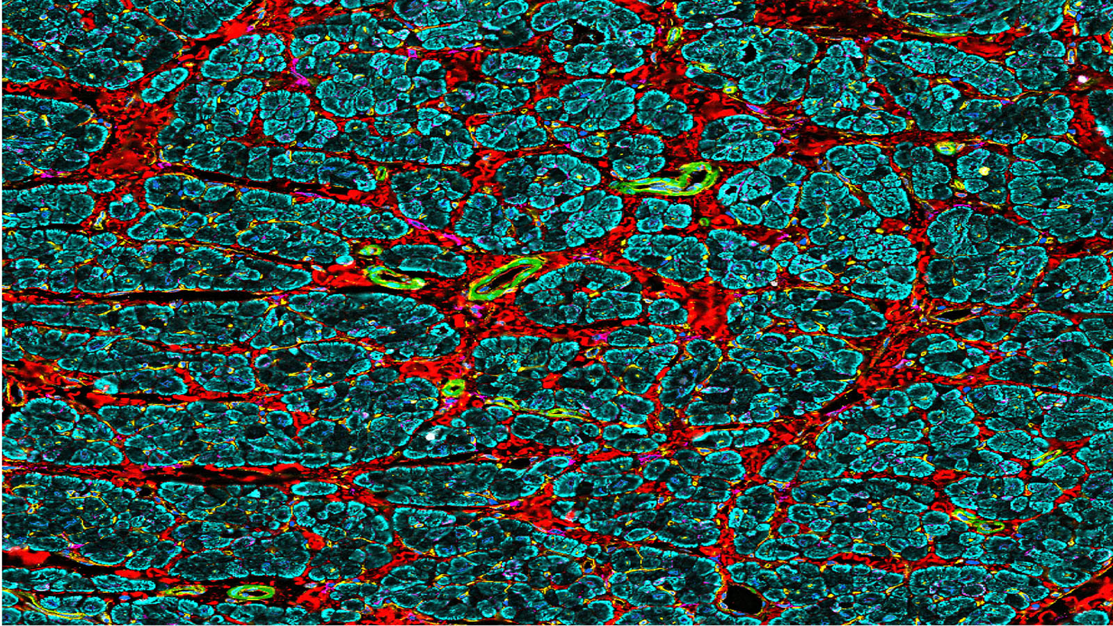
Confocal image of healthy human pancreas from Martha Campbell-Thompson at University of Florida
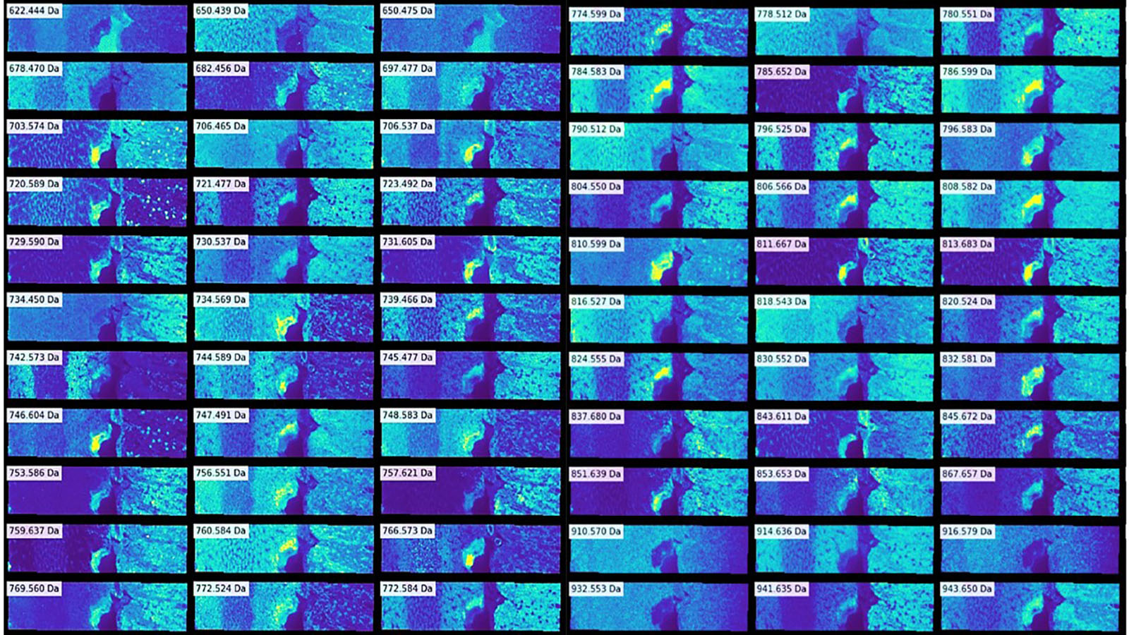
Mosaic of Imaging Mass Spec images of human kidney, courtesy of Dr. Elizabeth Neumann of Vanderbilt
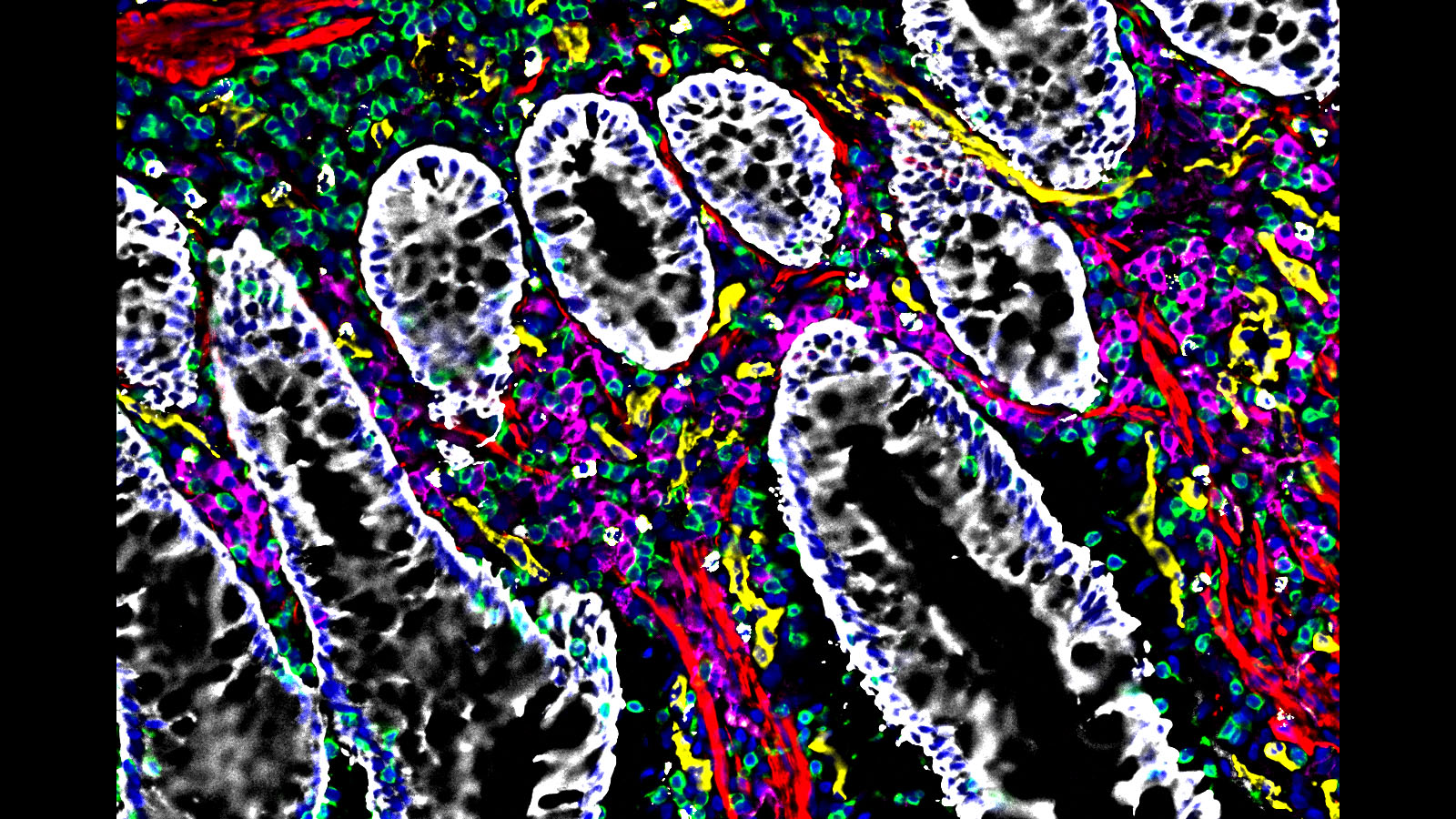
#CODEX image of the intestine courtesy of @stanford researcher Dr. John Hickey
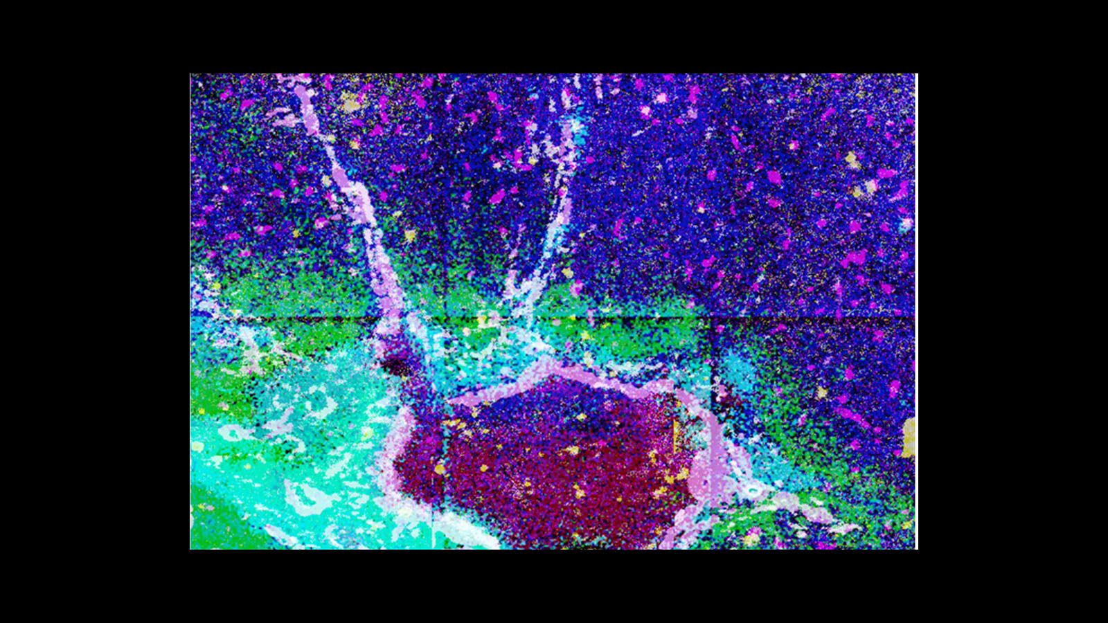
Integrated multiomics data from Imaging Mass Spec experiments from the liver, courtesy of Drs. Hua Tian (Penn State) and Brent Stockwell (Columbia University)
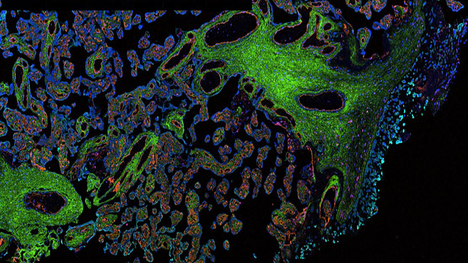
IMC image of placental trophoblasts courtesy of Dr. Santhosh Sivajothi of Jackson Labs
CODEX image of the left atrium of the heart from Dr. Kai Tan at CHOP
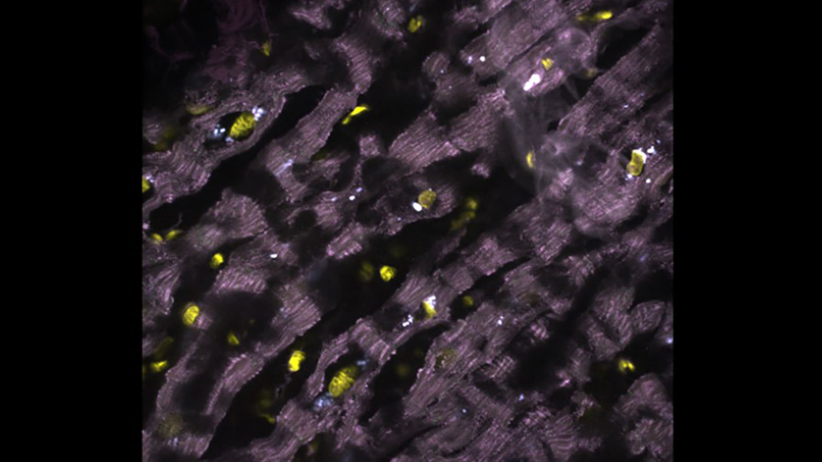
seqFISH image of healthy human heart, courtesy of Long Cai's lab at CalTech
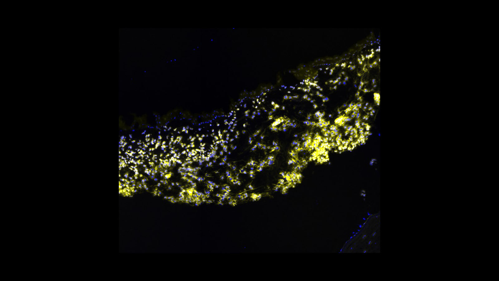
Fluorescent microscopy image of the human iris, courtesy of Dr. Angela Kruse from Vanderbilt
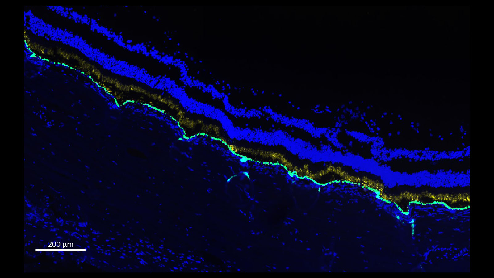
Fluorescent microscopy image of human retina, courtesy of Dr. Angela Kruse of Vanderbilt University
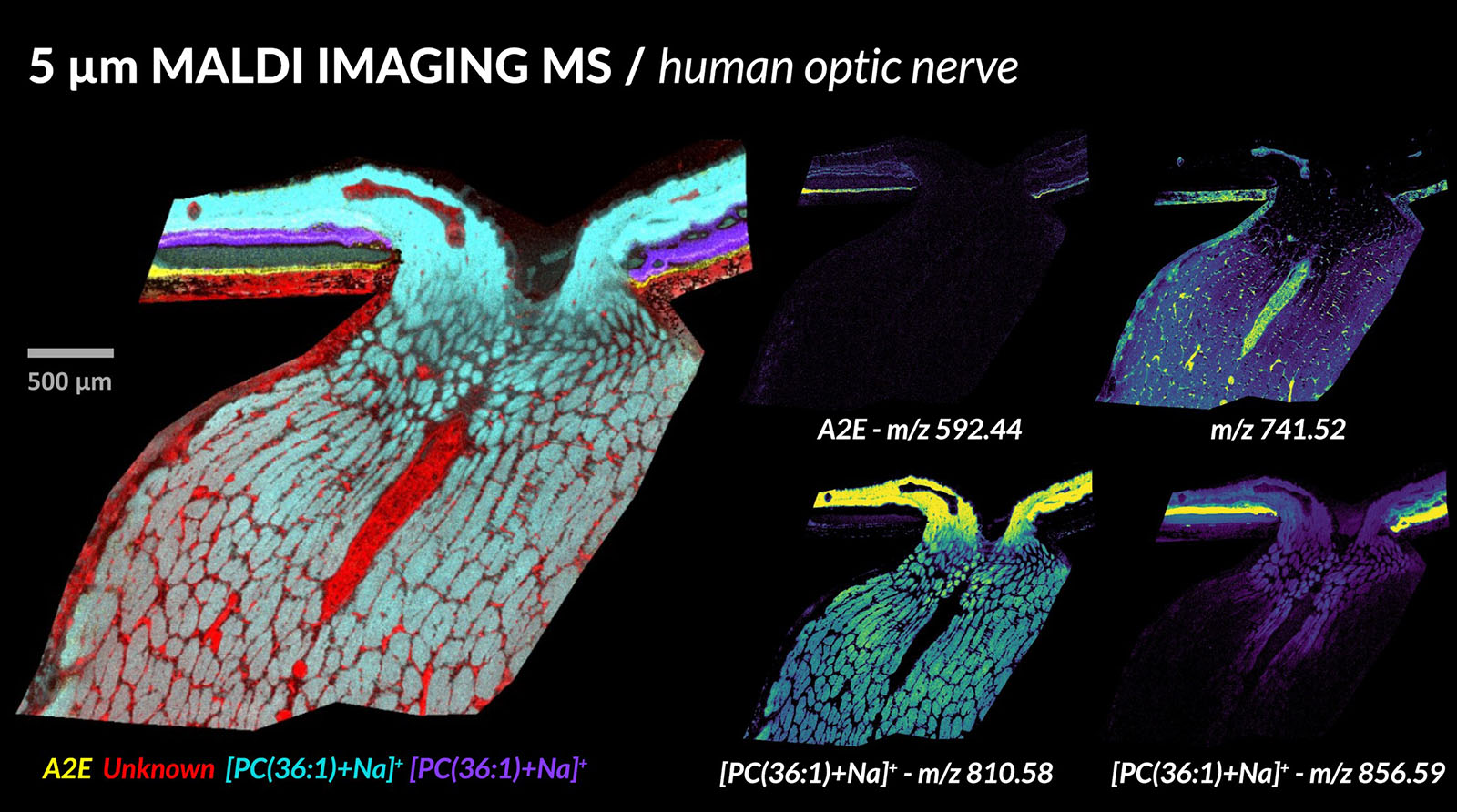
MALDI image of human optic nerve, courtesy of Dave Anderson from Vanderbilt
