
The Human BioMolecular Atlas Program (HuBMAP)
The Human BioMolecular Atlas Program (HuBMAP)
Image of the Week
Some of the most amazing things to come out of the HuBMAP Consortium are the images of healthy human tissues generated by our researchers.
Here, we collected them in one place to celebrate the work of these talented individuals.
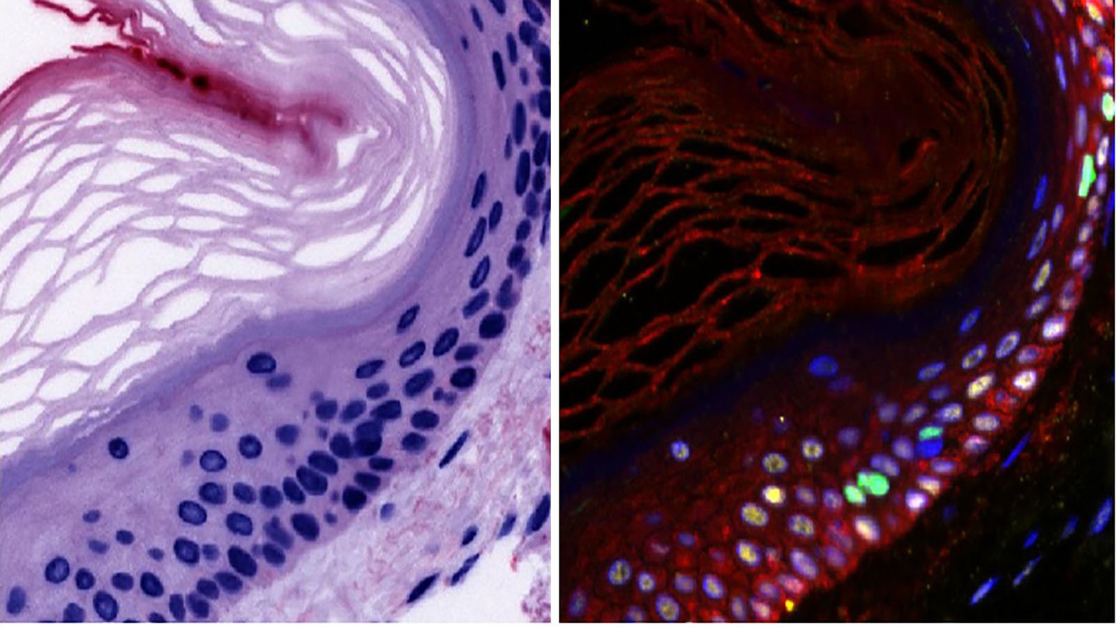
H&E and Cell Dive images of epidermis from Dr. Fiona Ginty at GE Research
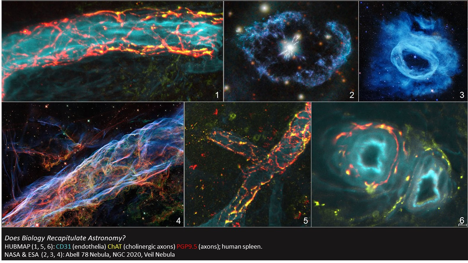
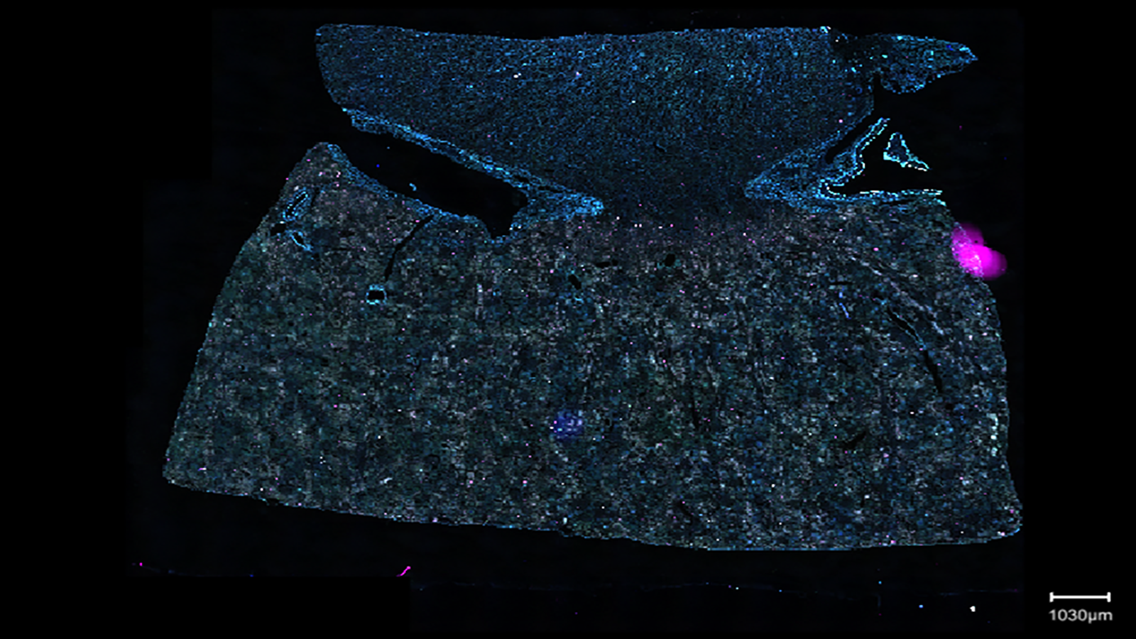
Lightsheet microscopy image of human spleen compared to nebula from Dr. Seth Currlin at University of Florida
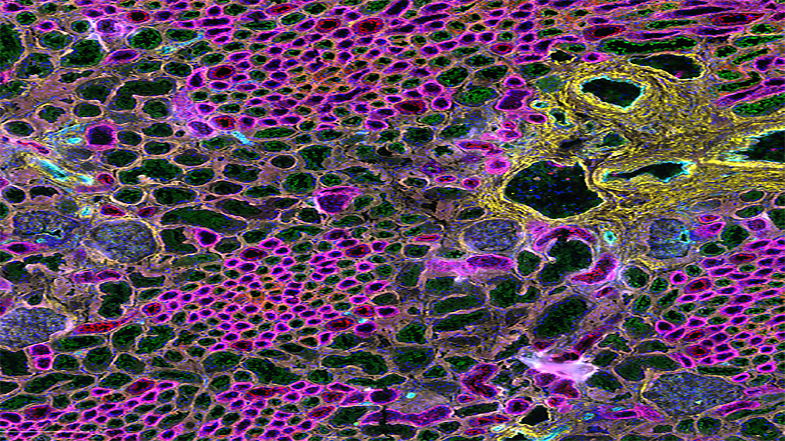
CODEX of healthy human colon, from Dr. John Hickey of the Nolan lab at Stanford
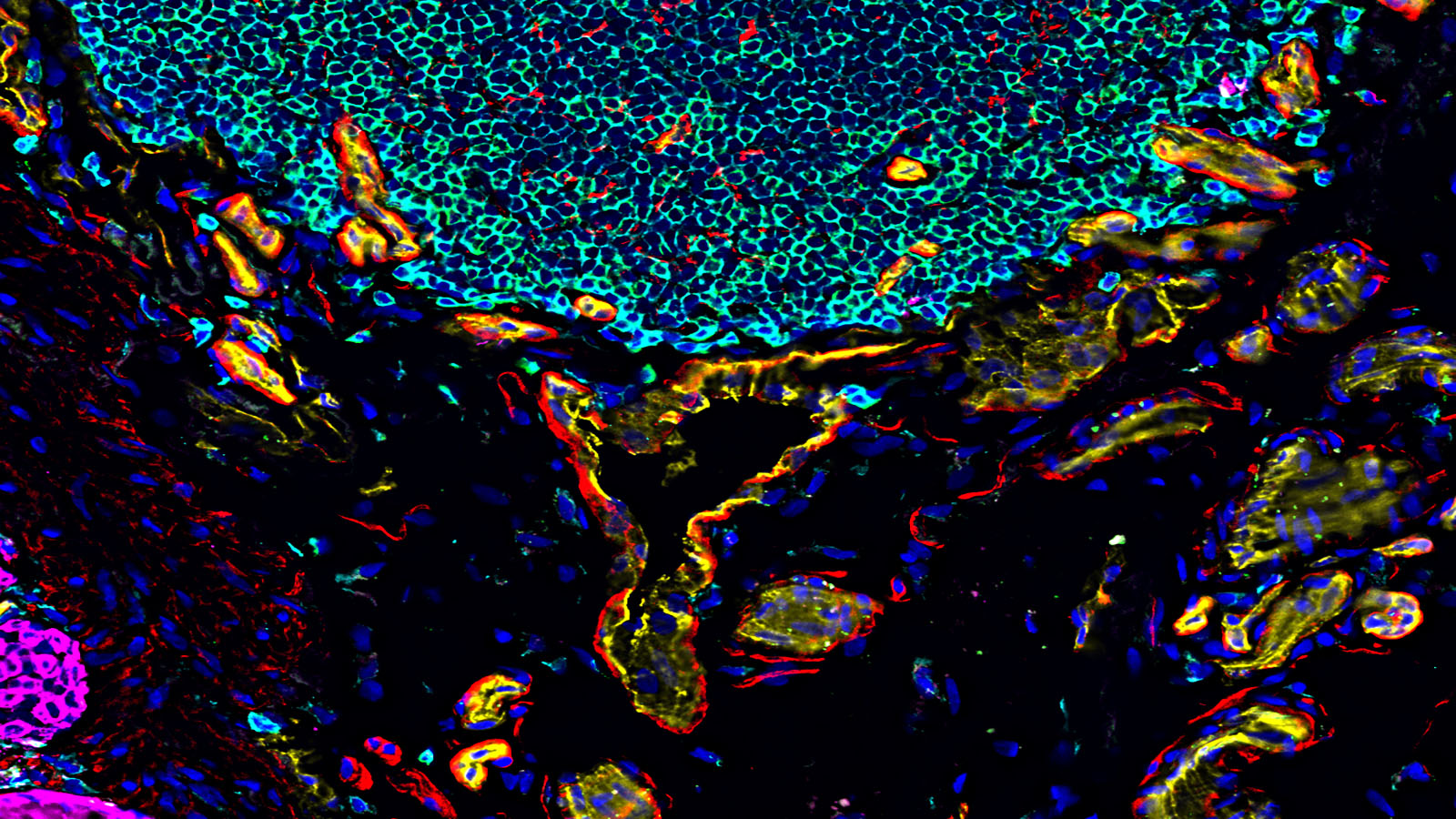
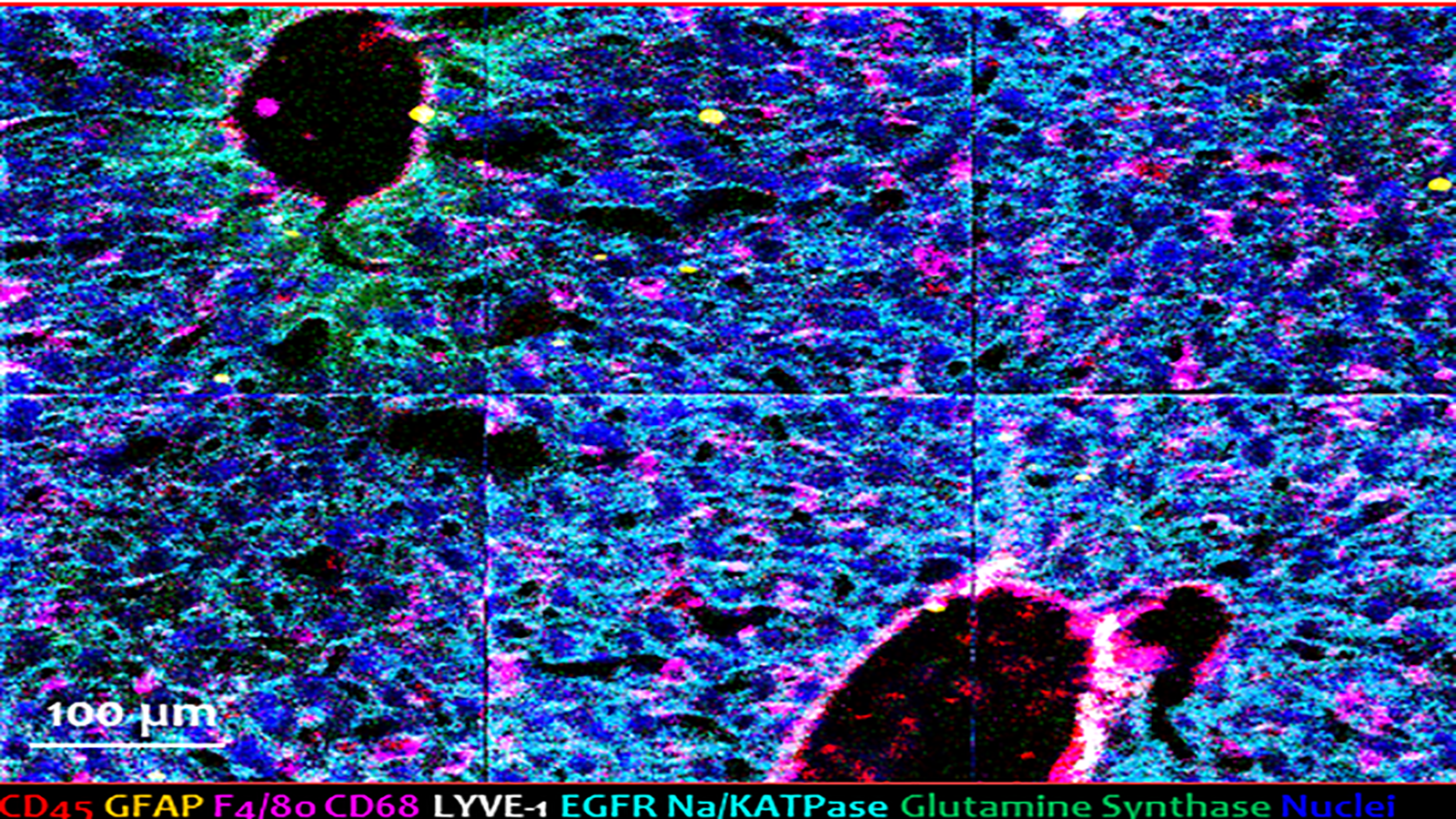
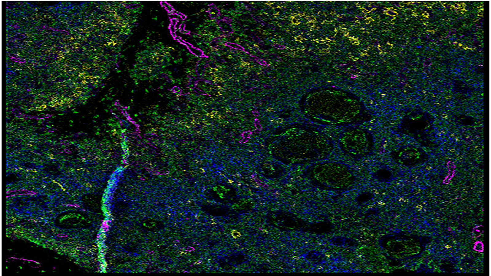
Cell DIVE image of healthy human kidney, courtesy of Christine Surrette and Dr. Elizabeth Neumann from GE Research and Vanderbilt (respectively)
Mass spec image of mouse liver, courtesy of Dr. Hua Tian at Penn State.
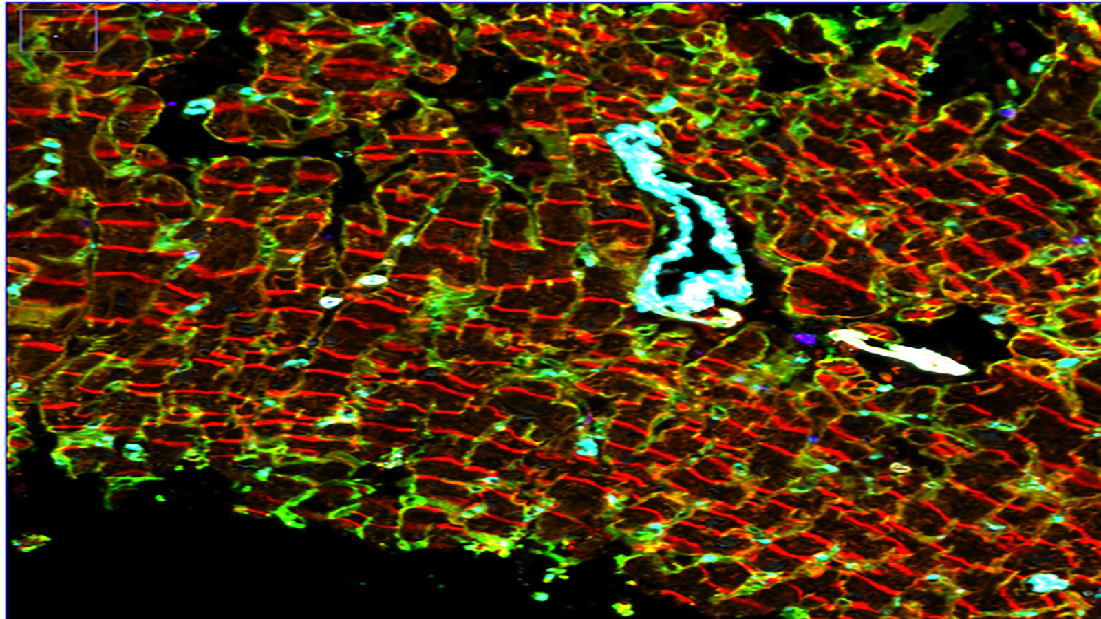
Cell DIVE image of the human heart, courtesy of Liz McDonough of the Ginty lab at GE Research
seqFISH image of healthy human small intestine, courtesy of Long Cai's lab at Cal Tech
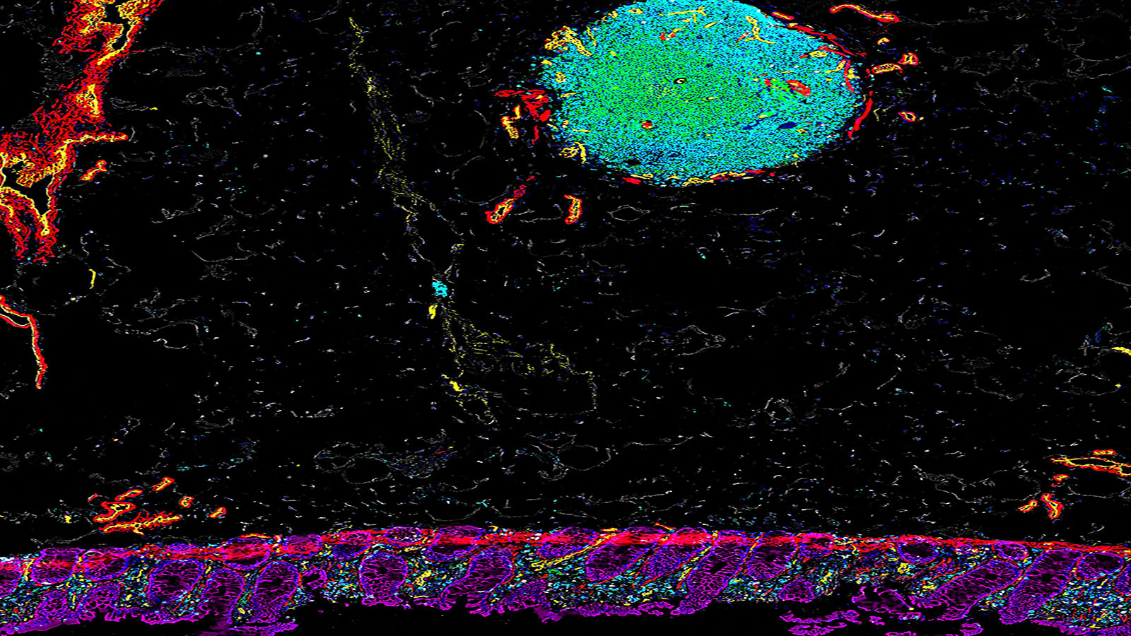
CODEX of healthy human colon, courtesy of Dr. John Hickey at Stanford
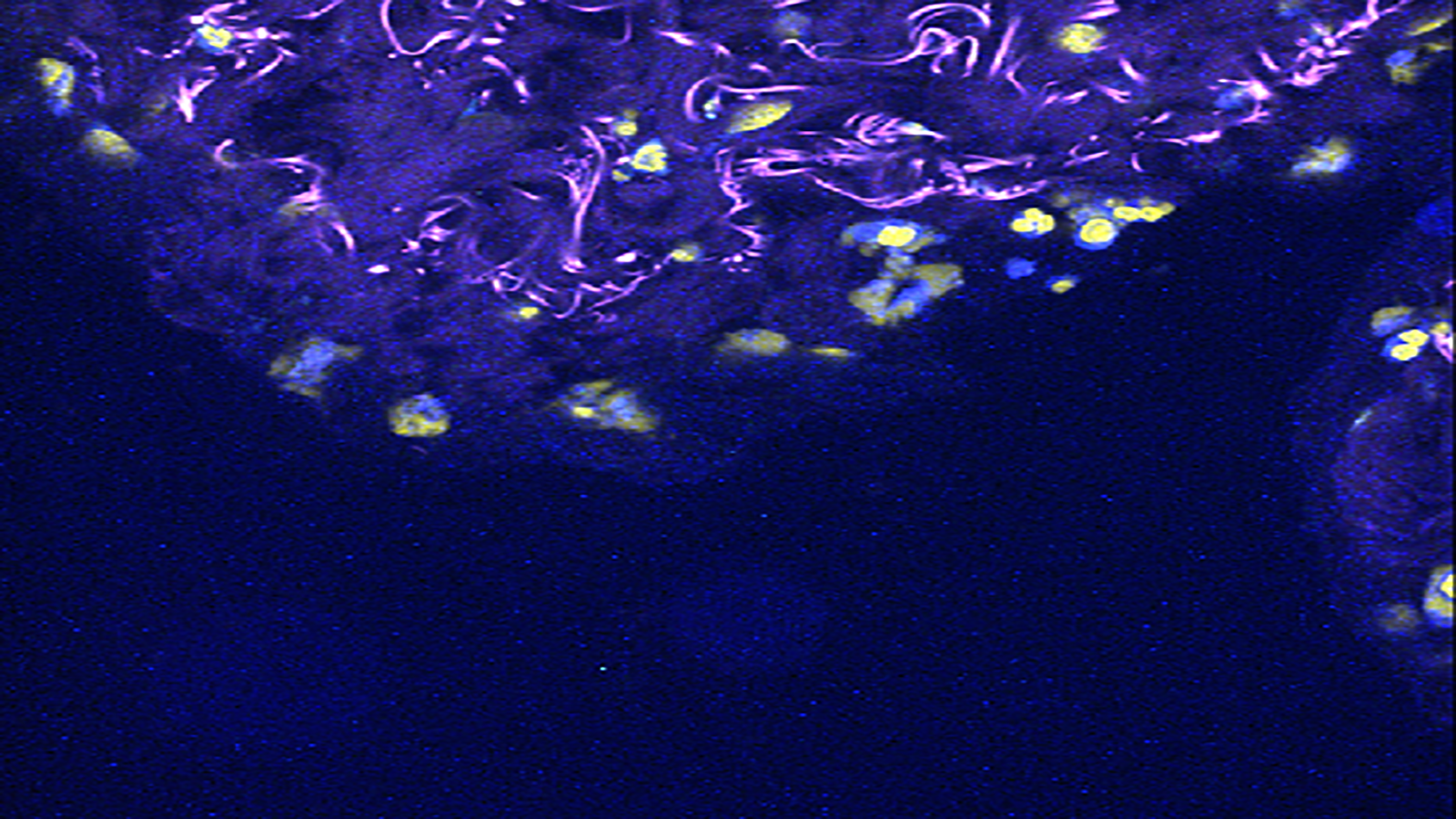
Imaging mass cytometry image of the thymus, courtesy of Michelle Daniel of the Bodenmiller Lab
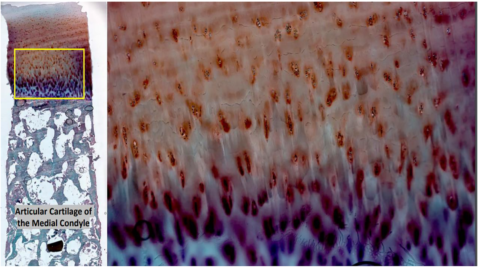
Human cartilage from the knee end of an adult femur stained with Safranin-O, courtesy of Dr. Peter Maye of UConn
Autofluorescence image showing the cortex, medulla, glomeruli, and proximal tubules of the human kidney, courtesy of Elizabeth Neumann at Vanderbilt
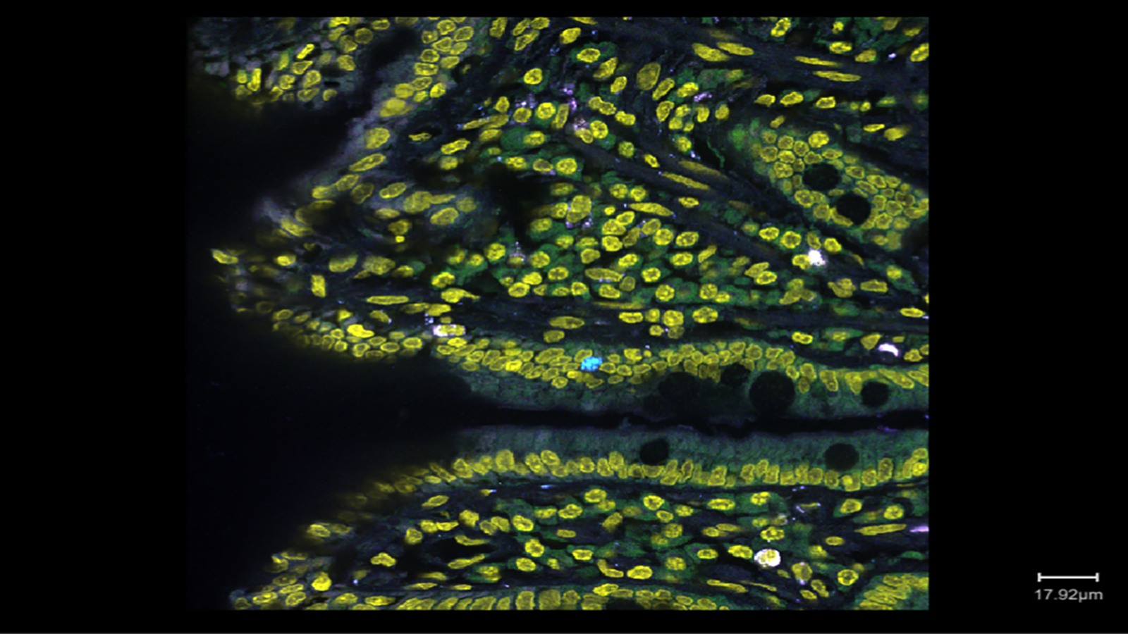
RNA transcripts in a section of healthy human small intestine from Long Cai's lab at Cal Tech
