
The Human BioMolecular Atlas Program (HuBMAP)
The Human BioMolecular Atlas Program (HuBMAP)
Image of the Week
Some of the most amazing things to come out of the HuBMAP Consortium are the images of healthy human tissues generated by our researchers.
Here, we collected them in one place to celebrate the work of these talented individuals.
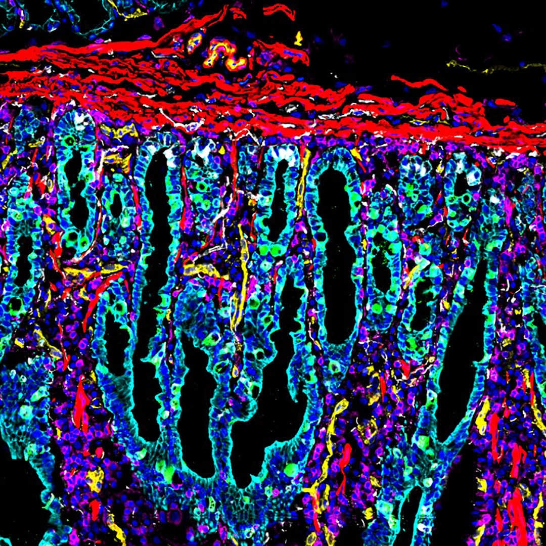
CODEX image of small intestine from Dr. John Hickey at Garry Nolan's lab at Stanford University
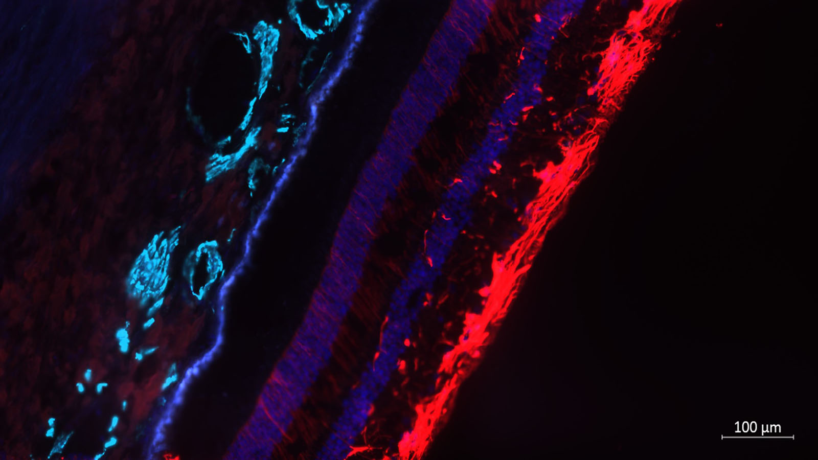
Image of a human retina from Dr. Angela Kruse at Vanderbilt
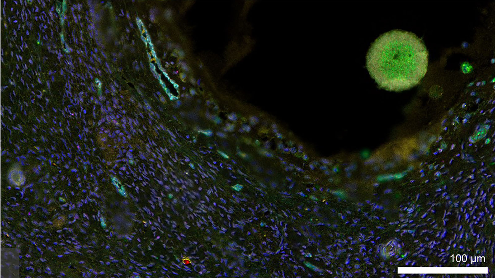
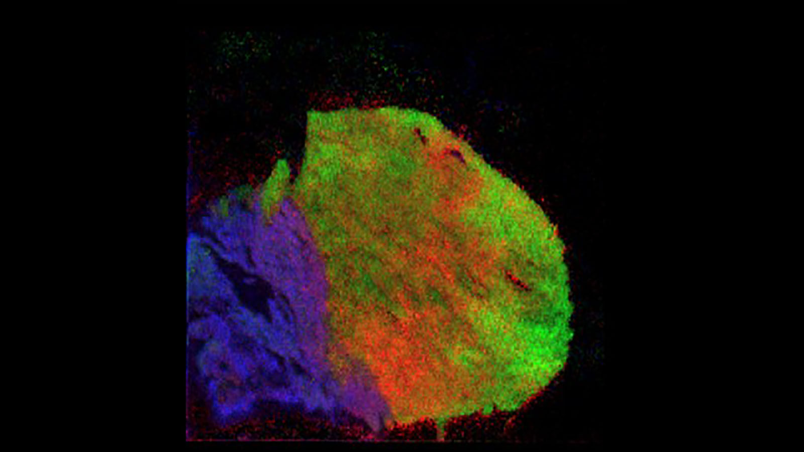
Microscopic image of an oocyte within its follicle, courtesy of Elizabeth Tsui in Dr. Monica Laronda's lab @Northwestern
MERFISH image of healthy human lung courtesy of Drs. Quan Shu, Colin Kern, Jamie Verheyden, and Xin Sun at UCSD, and Dr. Gloria Pryhuber at URMC
DESI image of mouse heart courtesy of Taruna Neelakantan from Columbia University
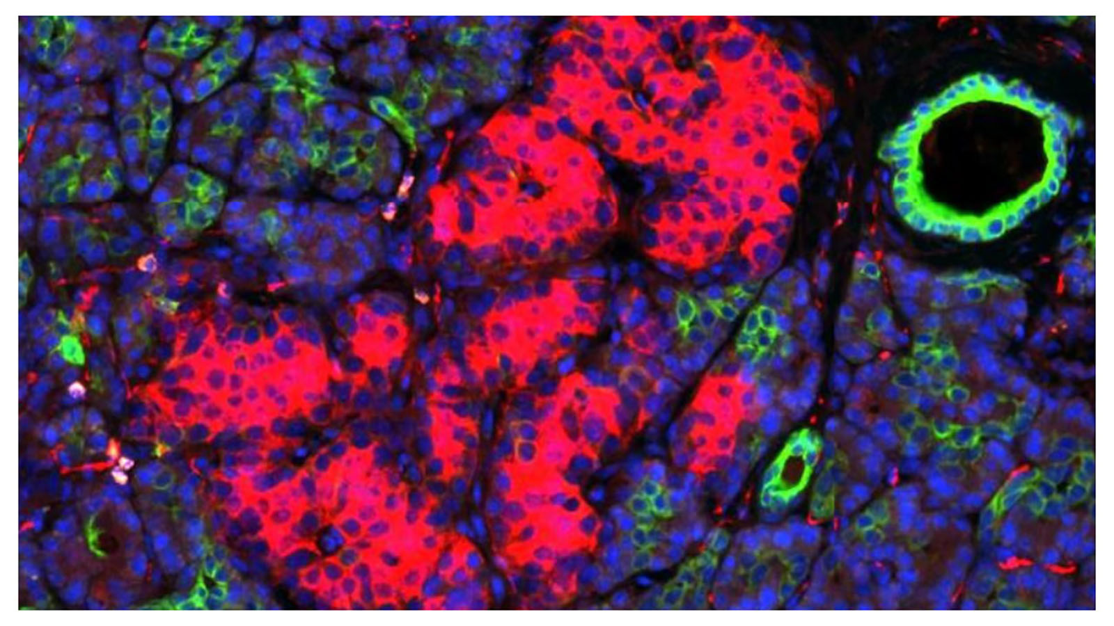
Nanostring GeoMX transcriptome image of human pancreas from Dr. Martha Campbell-Thomson at University of Florida
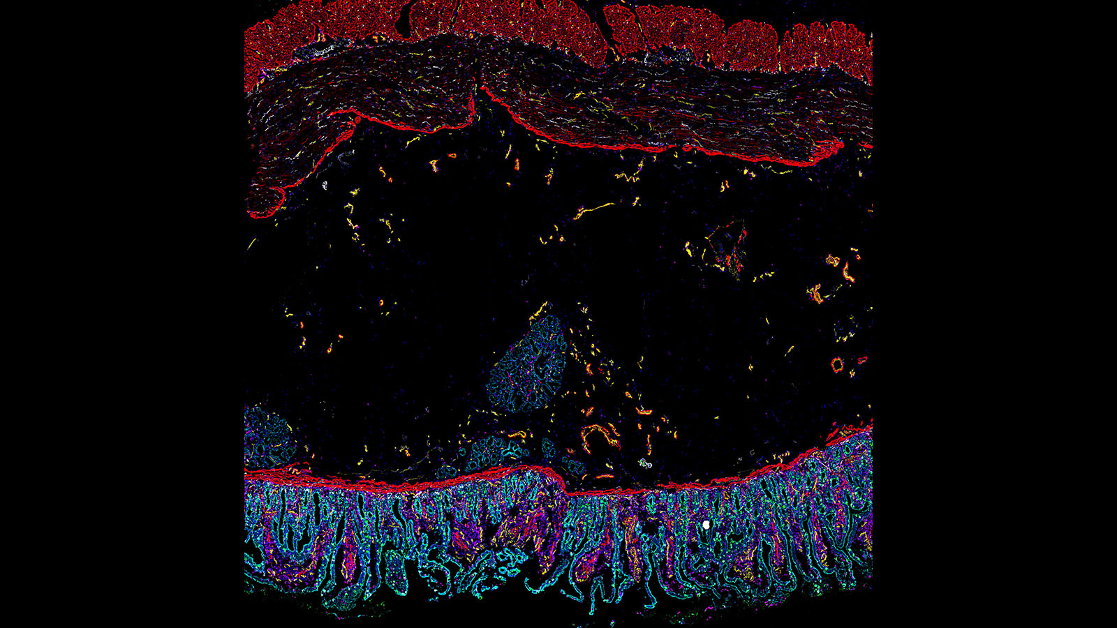
CODEX image of human intestine from Dr. John Hickey at Stanford University
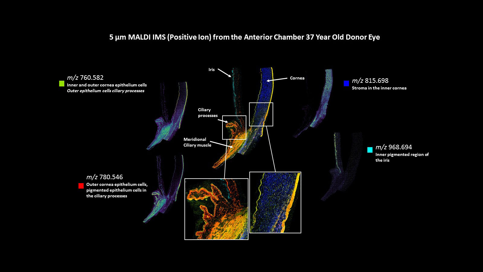
MALDI IMS image of the human cornea, courtesy of Dr. David Anderson from Vanderbilt University
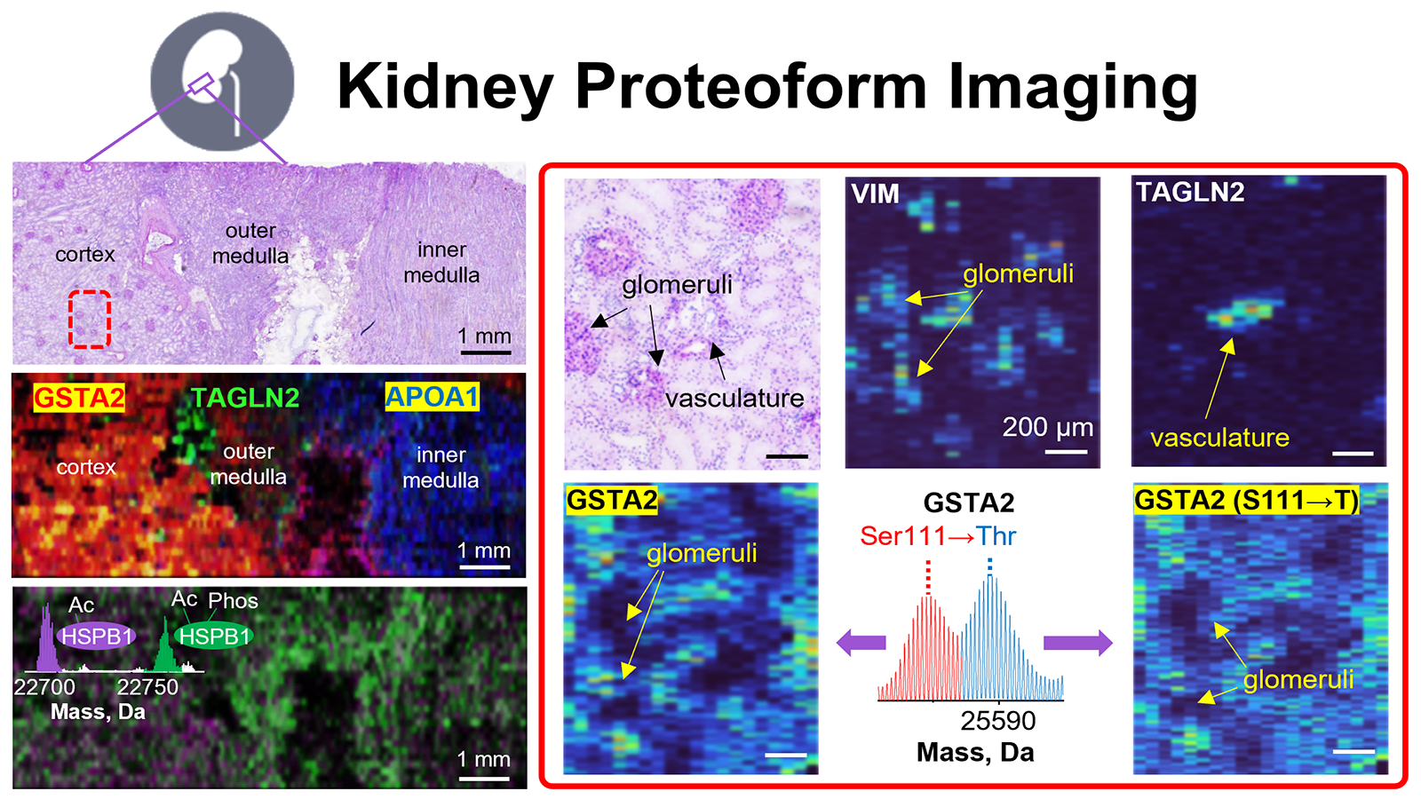
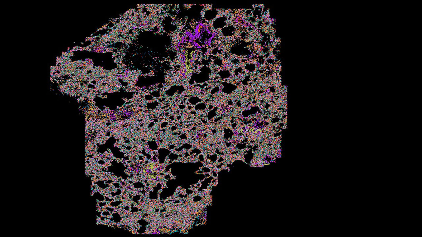
Proteoform imaging mass spec (PiMS) image of human kidney courtesy of Dr. Neil Kelleher at Northwestern
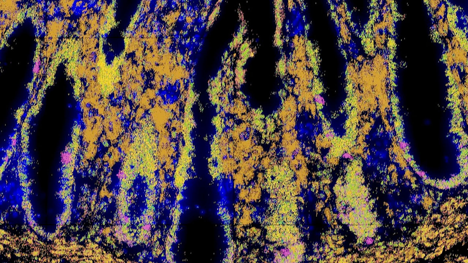
Molecular Cartography image of colon cells courtesy of Dr. Chenchen Zhu at Stanford
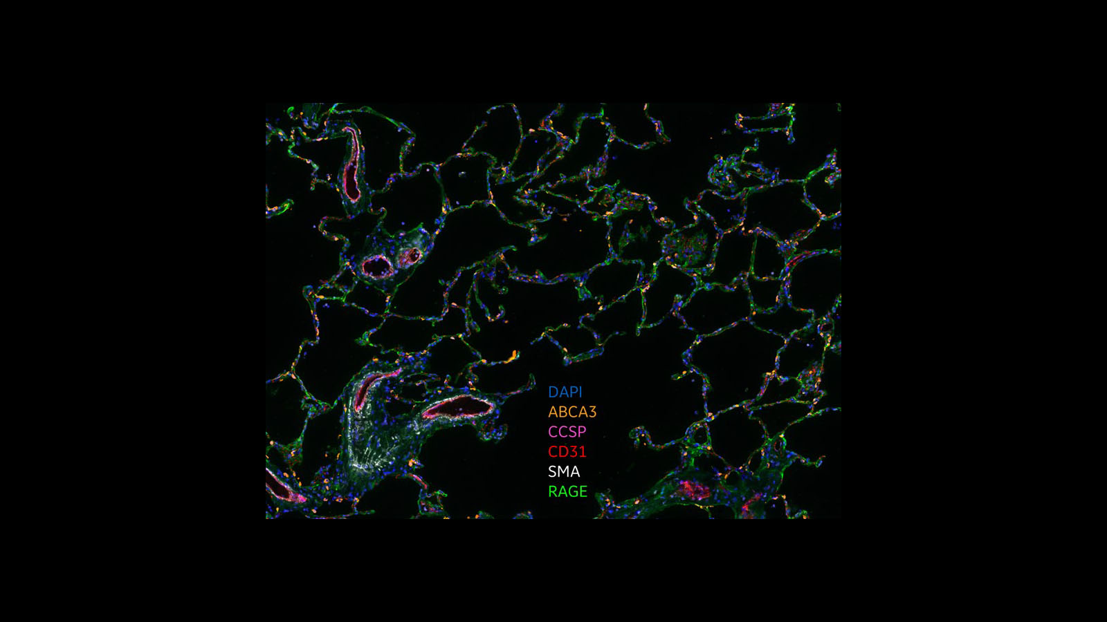
CellDIVE image of alveolar parenchyma cells from GE Research and University of Rochester
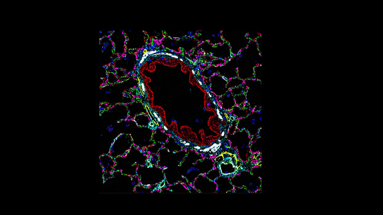
CODEX image of lung and small pulmonary arterial cells courtesy of Dr. Gloria Pryhuber
