
The Human BioMolecular Atlas Program (HuBMAP)
The Human BioMolecular Atlas Program (HuBMAP)
Image of the Week
Some of the most amazing things to come out of the HuBMAP Consortium are the images of healthy human tissues generated by our researchers.
Here, we collected them in one place to celebrate the work of these talented individuals.
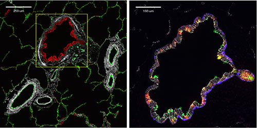
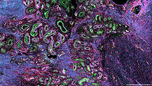
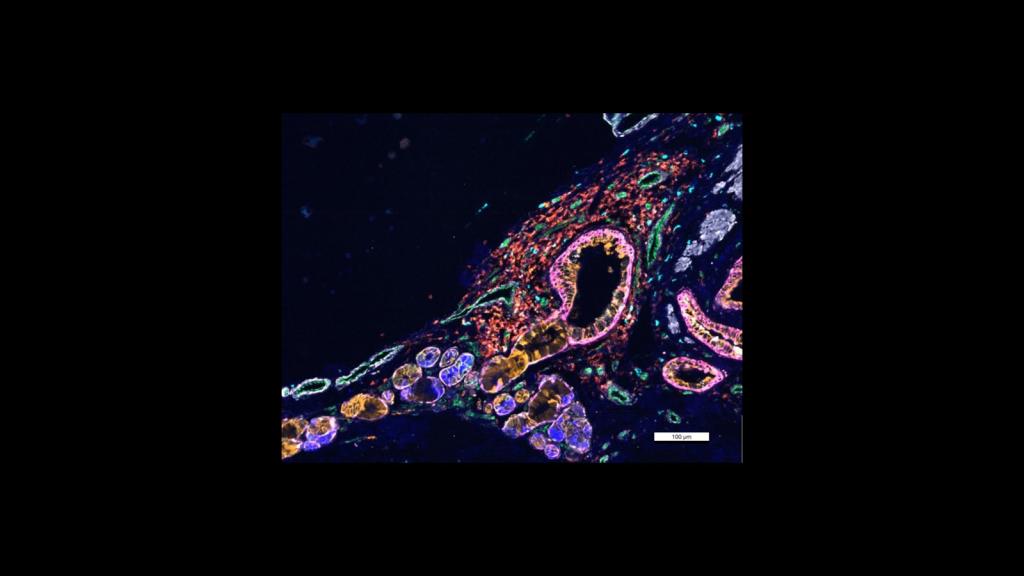
Two CODEX images of terminal bronchiole and associated vasculature, courtesy of Drs. Jeff Purkerson and Gloria Pryhuber at URMC
CODEX image of right atrium courtesy of Dr. Kai Tan at CHOP
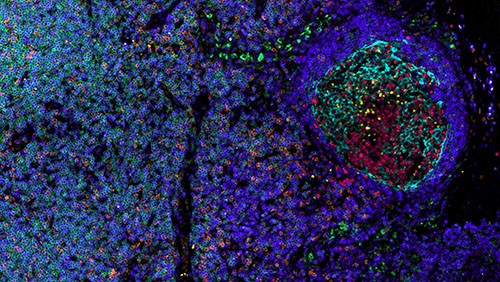
CellDIVE image of lymph node from Dr. Andrea Radtke in the Germain lab at NIAID
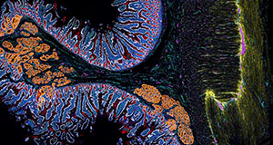
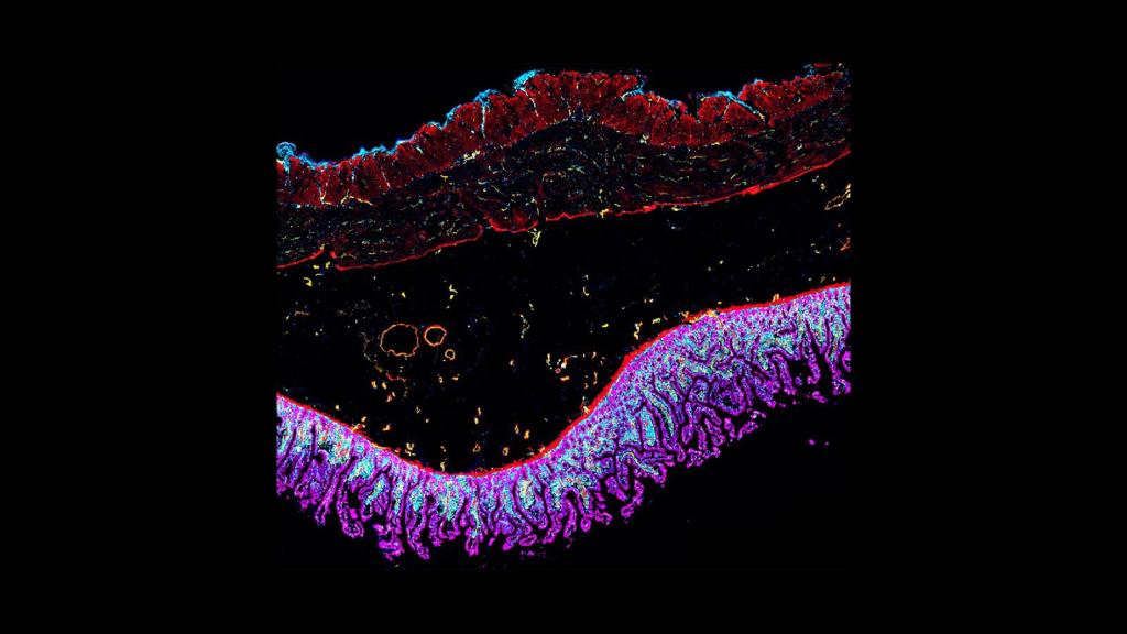
CODEX image of duodenum courtesy of Joanna Bi and Dr. Bei Wei of the Snyder lab at Stanford University
CODEX image of ovary from Elizabeth Tsui, Hannah McDowell from the Laronda lab at Lurie Childrens Hospital, and Alex Cabrera, Jean Rosario from the O'Neill lab at UPENN
CODEX image of lymph nodes, courtesy of Archie Enninful in the Fan lab at Yale
Cell DIVE image of DNA damage in skin cells from sun exposure courtesy of Dr. Liz McDonough at in the Ginty lab at GE Research
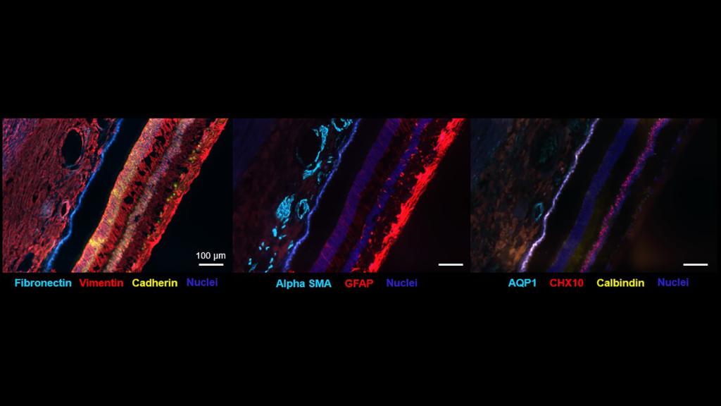
CODEX image of the human retina courtesy of Dr. Angela Kruse at Vanderbilt University
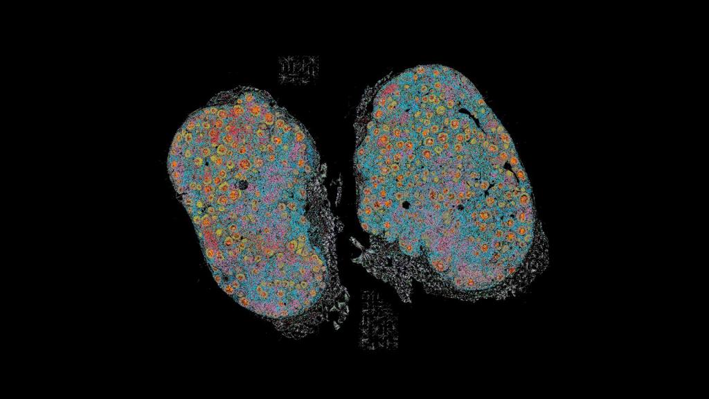
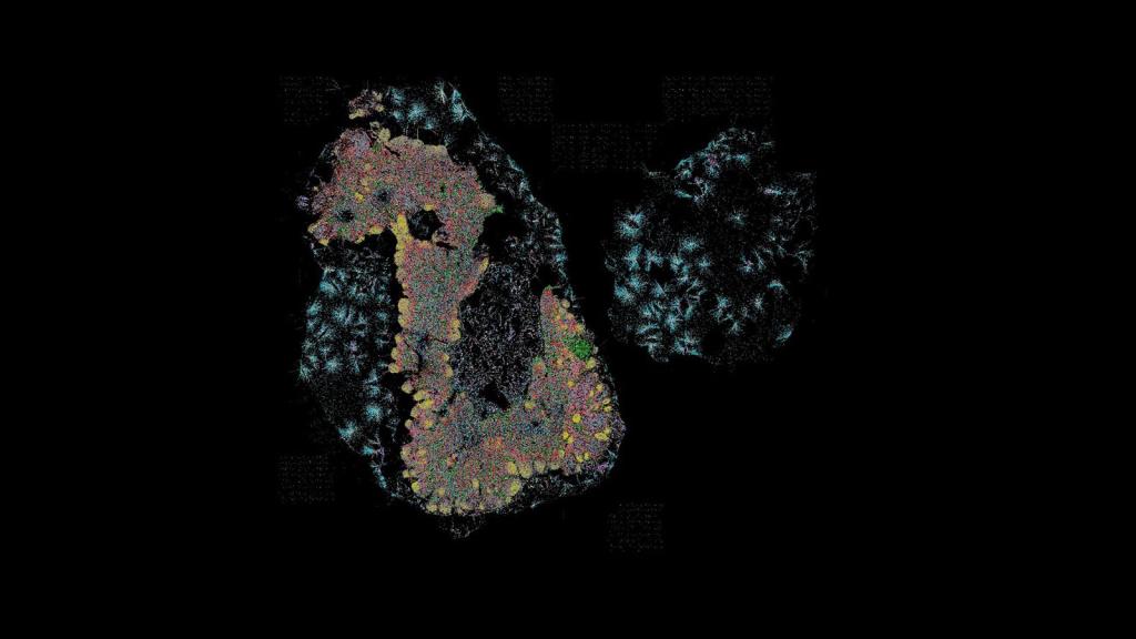
CODEX image of two lymph nodes, courtesy of Archie Enninful in Dr. Rong Fan's lab at Yale University
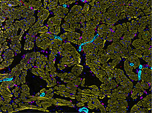
CellDIVE image of lung courtesy of Dr. Gloria Pryhuber at URMC
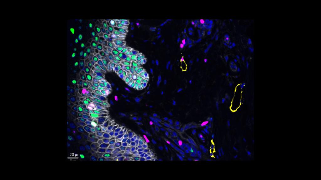
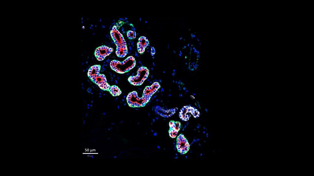
CODEX image of intestine courtesy of Dr. John Hickey from Stanford and Duke Universities
CellDIVE image of glands in skin, courtesy of Dr. Liz McDonough from GE
